If you have been researching hair loss or scalp conditions for any length of time, you have probably encountered the word trichoscopy. It appears in dermatology articles, in clinical guidelines, and increasingly in the context of AI-powered scalp analysis. But what it actually is — what it reveals, how it works, and why it matters for someone managing a scalp concern without access to a clinical setting — is rarely explained with precision.
This is that explanation.
What Trichoscopy Is
Trichoscopy is the application of dermoscopy — a technique that uses magnified, polarised light to examine skin structures — specifically to the scalp and hair. First formally described in the early 2000s, it has since become the standard non-invasive assessment tool for scalp and hair disorders across trichology and dermatology practice globally.1
A trichoscope illuminates the scalp at high magnification using multiple light modes simultaneously. Standard white light reveals surface structures: scaling patterns, follicular openings, hair shaft morphology, and vascular patterns visible at the scalp surface. Polarised light penetrates the upper epidermal layers, revealing subsurface inflammation, pigmentary changes, and structural detail that no surface examination — clinical or otherwise — can detect. UV light at 365nm makes bacterial colonisation visible as fluorescent porphyrin deposits, a diagnostic detail relevant to conditions including seborrheic dermatitis that no other non-invasive assessment method identifies reliably.
What Trichoscopy Reveals
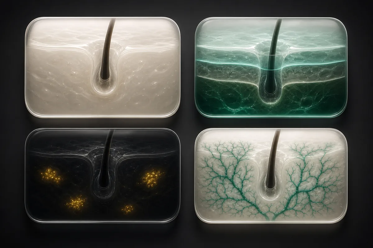
The clinical value of trichoscopy lies in what it makes visible at the follicle and scalp structure level — indicators that are measurable, documentable, and trackable across time with serial assessments.
Follicle density and miniaturisation are among the most diagnostically significant findings in trichoscopic assessment. In androgenetic alopecia, DHT-sensitive follicles undergo progressive miniaturisation — hair shaft diameter narrows across successive growth cycles until the follicle produces only fine vellus hair. This process is measurable under trichoscopy weeks to months before it produces any visible density change at the scalp surface.2 Trichoscopic imaging can detect the early shaft diameter variability that marks the beginning of follicle sensitisation — the trichoscopic signature of androgenetic alopecia — before it is apparent to any other non-invasive assessment method.
Scalp inflammation markers — perifollicular scaling, interfollicular scaling, and the distribution pattern of inflammatory change around follicular openings — are reliably identified under trichoscopy and correlate with conditions including seborrheic dermatitis, psoriasis, and lichen planopilaris. The pattern of inflammation, not merely its presence, is what guides clinical differentiation between these conditions.
Hair shaft abnormalities — shaft diameter variability, circle hairs, coil hairs, shaft fractures, and taper failures — are characteristic findings in specific alopecia patterns. Their distribution across scalp zones, and their evolution over time with treatment, is documentable through serial trichoscopy in a way that no other non-invasive method supports.3
Vascular patterns at the scalp surface — simple loops, arborising vessels, and glomerular vessels — correlate with specific inflammatory and non-inflammatory conditions and are assessed as standard elements of a complete trichoscopic evaluation.
What Trichoscopy Cannot Do
This is the point where clinical precision matters most — and where accurate language is more useful than reassuring overclaim.
Trichoscopy identifies patterns that may be associated with specific scalp and hair conditions. It does not diagnose. A finding of significant follicle miniaturisation with perifollicular pigmentation in specific scalp zones may be strongly associated with androgenetic alopecia — but the clinical picture, patient history, hormonal context, and full differential diagnosis are what a qualified clinician, such as a dermatologist, brings to the interpretation of those findings. The trichoscope produces evidence. The clinician interprets it.
This distinction is not a limitation — it is the correct description of how evidence-based clinical assessment works. Understanding it accurately is more useful, and more trustworthy, than any claim that a technology can determine a diagnosis independently of clinical judgment.
Why This Matters Specifically for Postpartum Hair Loss
Postpartum telogen effluvium and early-stage androgenetic alopecia can present similarly in the months following delivery — particularly at the frontal hairline and temples, where both conditions produce visible change. The distinction between them has significant clinical implications: telogen effluvium typically resolves spontaneously within a defined biological window; androgenetic alopecia requires a different clinical pathway and responds to different interventions.
Under trichoscopy, the two conditions produce different findings. Telogen effluvium typically shows increased numbers of empty follicular openings — the telogen unit — with preserved shaft diameter and absent miniaturisation. Early androgenetic alopecia shows diameter variability: a mixture of terminal hairs and miniaturising hairs within the same zone, which is the trichoscopic signature of DHT-mediated follicle sensitisation.4 This distinction is measurable at the follicle level. It is invisible to visual self-assessment and indistinguishable by casual photography.
Our complete guide to why hair thins after pregnancy covers both the biology of postpartum telogen effluvium and the clinical signals that indicate when the pattern warrants professional assessment rather than watchful waiting.
Where Scalp Score Fits
Scalp Score by Orga Hair is not trichoscopy, and it does not try to be. There is no instrument, no polarised or UV light and no magnification — an app on your phone cannot see what a trichoscope sees. What it can do is help you keep track of your own scalp, wherever you are.
It starts with five short questions and gives you a Scalp Score straight away, worked out on your phone with no sign-up. The score is an estimate built from your answers, not a measurement. Free tools sit around it: a routine built around your wash days, a wash-day check-in, the Product Checker, and a Photo Journal that keeps your own photographs in order so you can compare them later.
When you want to look closer, Scalp Score Pro is a one-off purchase in Google Play: an AI analysis of the photographs your answers call for — normally two or three scalp zones — read together with your profile. It describes what your photographs show in a five-part report (Today, Where, Why, Plan, Watch) and says what it could not assess. If a report or your own record raises a question, a GP or dermatologist is the person to take it to — and trichoscopy is one of the tools they may use.
Who Benefits Most from a Trichoscopic Baseline
A clinical trichoscopy assessment is most valuable at specific moments in a hair and scalp health journey.
It is most valuable at the point of concern, before any treatment or protocol is started. A baseline at week zero establishes the reference point against which all subsequent measurement is compared. Without it, as documented in our article on the risks of self-treating female hair loss, it is not possible to assess whether any intervention is producing improvement, has plateaued, or is masking subclinical regression behind other variables.
It is valuable during active treatment, at defined intervals — weeks 4, 8, and 12 — to assess whether the trajectory is moving in the expected direction. Trichoscopy used longitudinally at these intervals has been validated as a sensitive method for monitoring treatment response in androgenetic alopecia, detecting subclinical follicle activity before it becomes visible at the surface level.5
And it is valuable before a clinical appointment — not as a replacement for it, but as the preparation that transforms a first consultation from a starting point into a structured, evidence-based conversation. The documented resolution advantage of professional diagnosis over self-directed treatment begins with arriving at the appointment with a structured clinical picture rather than a history reconstructed from memory.
If a trichoscopy appointment is not within reach yet, you can still start a record of your own. Scalp Score gives you a free starting point from five short questions, and its Photo Journal and wash-day check-ins help you keep track of what changes — so that if you do see a GP or dermatologist, you arrive with more than memory.
References
- Rudnicka L, Olszewska M, Rakowska A, et al. (2008). Trichoscopy: a new method for diagnosing hair loss. Journal of Drugs in Dermatology, 7(7), 651–654. Read study
- Rakowska A, Slowinska M, Kowalska-Oledzka E, et al. (2009). Dermoscopy in female androgenic alopecia: method standardization and diagnostic criteria. International Journal of Trichology, 1(2), 123–130. Read study
- Rudnicka L, Rakowska A, Kerzeja M, & Olszewska M. (2013). Hair shafts in trichoscopy: clues for diagnosis of hair and scalp diseases. Dermatologic Clinics, 31(4), 695–708. Read study
- Miteva M, & Tosti A. (2012). Hair and scalp dermoscopy. Journal of the American Academy of Dermatology, 67(5), 1040–1048. Read study
- Blume-Peytavi U, Hillmann K, Dietz E, et al. (2011). A randomized, single-blind trial of 5% minoxidil foam once daily versus 2% minoxidil solution twice daily in the treatment of androgenetic alopecia in women. Journal of the American Academy of Dermatology, 65(6), 1126–1134. Read study
- Olszewska M, Rudnicka L, Rakowska A, et al. (2012). Use of dermoscopy in trichology. Journal of Dermatological Case Reports, 3(2), 14–20. Read study