
The shedding can start months after the thyroid event that caused it — sometimes so long after that the two feel entirely unconnected. The follicle does not immediately shed when thyroid function changes. It first shifts through the stages of its own cycle, and the visible hair loss arrives only after that process completes. By then, the patient is typically looking for a cause in the present rather than in what was happening three or four months earlier.
This timing gap — biological, well-documented, and persistently underrecognised — is one of the reasons thyroid-related hair loss remains one of the more commonly missed factors in diffuse shedding presentations. Understanding why the delay exists, what TSH actually measures, and when a fuller thyroid panel adds clinical context changes the quality of the conversation that is possible with a GP or endocrinologist.
Two directions, one diffuse pattern
Both an underactive thyroid (hypothyroidism) and an overactive thyroid (hyperthyroidism) can drive diffuse hair shedding through telogen effluvium. Thyroid hormones — primarily T3 (triiodothyronine) and T4 (thyroxine) — directly regulate hair follicle activity, and when their levels are significantly disrupted in either direction, follicles may exit the active anagen growth phase prematurely and accumulate in the resting telogen phase.
The severity, however, is not equivalent between the two directions. A retrospective study of 500 women with confirmed telogen effluvium by Bin Dayel and colleagues found that the hypothyroid group had significantly higher mean SALT (Severity of Alopecia Tool) scores than both the euthyroid and hyperthyroid groups, with 34% of hypothyroid patients meeting the threshold for severe hair loss compared to 18% in the euthyroid group.3 The mean SALT scores for hyperthyroid patients did not differ significantly from those without thyroid dysfunction in the same cohort. Both directions are clinically relevant — but hypothyroidism, in this dataset, carries the heavier burden of shedding severity.
A broader systematic review by Hussein, Atia, and Bin Dayel confirmed that hyperthyroidism, hypothyroidism, and drug-induced hypothyroidism can all induce widespread hair shedding, and that the impact of thyroid disorders on hair problems remains underestimated in clinical practice.2
What thyroid hormones are doing inside the follicle
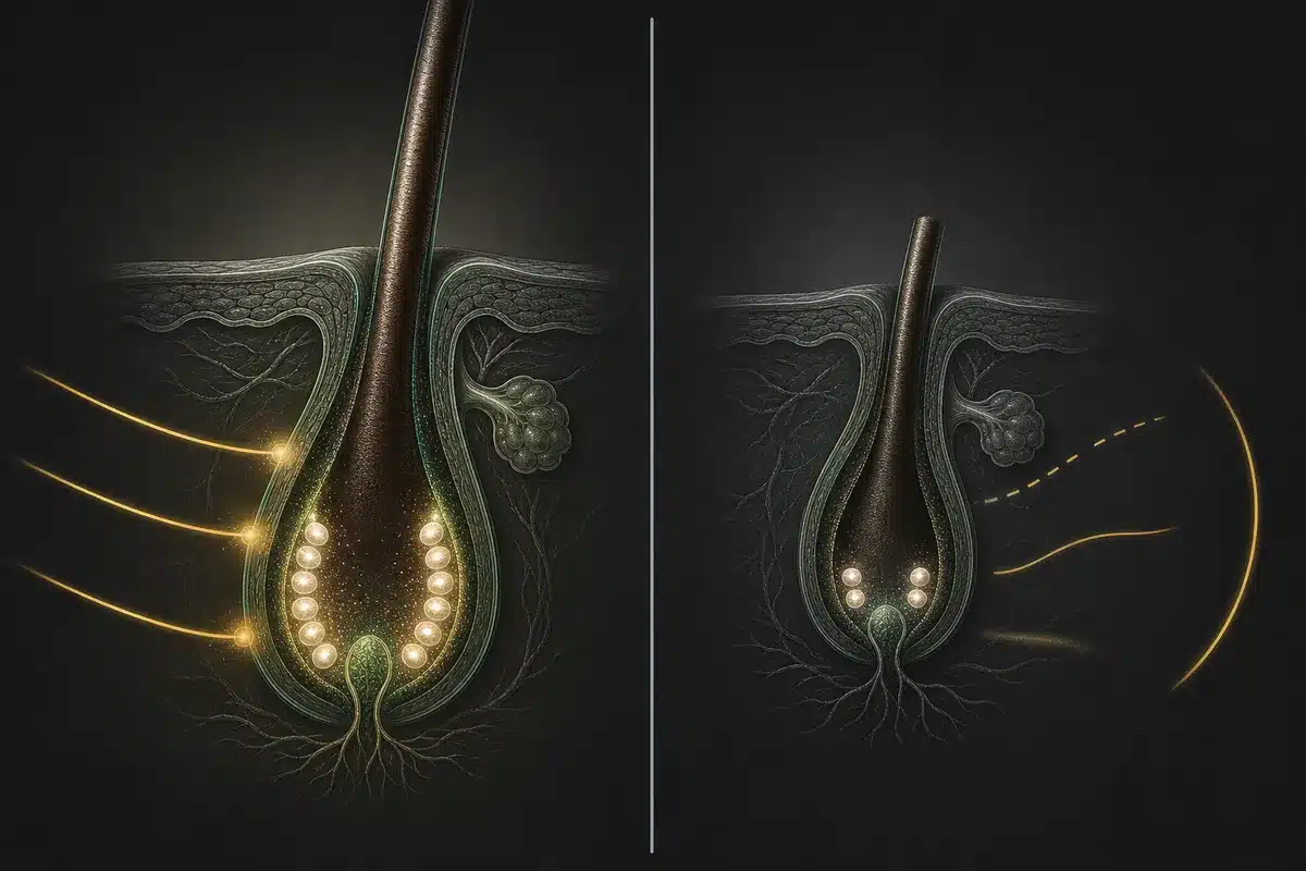
The relationship between thyroid hormones and the hair follicle is not simply indirect — that is, not merely a downstream consequence of altered metabolism or circulation. In 2008, van Beek and colleagues provided the first direct evidence that human scalp hair follicles are biological targets of T3 and T4, with their own capacity to respond to and process thyroid hormone signals.1
Their research, conducted on organ-cultured human scalp follicles from euthyroid women, found that T4 upregulates the proliferation of hair matrix keratinocytes — the rapidly dividing cells at the base of the follicle that produce the hair shaft. T3 and T4 together reduce the rate of matrix cell apoptosis, supporting the survival of actively growing follicle cells. Critically, T4 was shown to prolong the duration of the anagen phase, acting in part by downregulating TGF-β2 — a protein that functions as a key anagen-inhibitory growth factor, the biological signal that normally instructs the follicle to exit growth and enter regression.
The follicle also expresses the deiodinase enzymes D2 and D3, allowing it to convert T4 to the more biologically active T3 locally, within the follicle environment itself. When thyroid hormone levels fall below what the follicle’s own signalling requires, anagen maintenance loses direct biological support. When they are elevated, the precise hormonal timing governing cycle transitions is disrupted. In either case, the follicle has physiological reason to transition toward catagen and telogen earlier than its natural cycle would otherwise dictate.
The lag that makes it hard to see
Telogen effluvium does not appear at the moment of its trigger. The hair follicle first transitions from anagen to telogen — a process that itself takes several weeks — and the resulting telogen hair then sheds approximately two to three months after the triggering event. For thyroid-related TE, this means the diffuse shedding that presents in a clinic, in the shower drain, or on the brush arrived months after the thyroid disruption that caused it.
This creates a diagnostic mismatch that affects both recognition and recovery. A patient experiencing significant diffuse shedding in October is unlikely to connect it to thyroid changes that were occurring in the summer. Similarly, a patient whose thyroid function has been stabilised through treatment may expect their hair to recover promptly — and feel discouraged when shedding continues well into the months following normalisation of TSH. The delay in recovery follows the same biological logic: follicles that entered telogen before treatment began must complete that resting phase and re-enter anagen before new growth can resume. That transition, for each affected follicle, can take three to six months after hormone levels stabilise.
This also explains why a single TSH test taken at any one moment gives a limited picture. TSH can change over time — in response to illness, stress, nutritional shifts, pregnancy, or underlying disease progression — and a historical result does not establish what thyroid function looks like now, or how it has shifted relative to a previous baseline.
What TSH actually measures — and where free hormones and antibodies add context
TSH — thyroid-stimulating hormone — is produced by the pituitary gland, not by the thyroid itself. It functions as a feedback signal: when circulating thyroid hormone levels are insufficient, the pituitary releases more TSH to drive greater thyroid output. When thyroid hormones are excessive, TSH is suppressed. Elevated TSH is the earliest and most sensitive indicator of hypothyroidism; suppressed TSH points toward hyperthyroidism. For most clinical presentations of diffuse hair loss, TSH is the appropriate first-line test.
Its limits are also clinically meaningful. TSH reflects the pituitary’s current assessment of thyroid hormone availability — not the local hormonal environment at the tissue level. In subclinical thyroid dysfunction, TSH may be mildly elevated or suppressed while free T4 remains within its reference range, and clinical symptoms including hair changes may already be present. Free T4 and free T3 — the biologically active fractions of thyroid hormones not bound to carrier proteins — and thyroid antibodies (thyroid peroxidase antibodies, TPO-Ab; thyroglobulin antibodies, TgAb) can provide important additional context in selected clinical presentations. These decisions belong with a GP or endocrinologist, who can weigh the panel results alongside a full clinical history, symptom pattern, and relevant family history.
The autoimmune layer
For a subset of patients, the connection between thyroid health and hair loss involves a third pathway: shared immunological territory between autoimmune thyroid disease and autoimmune hair loss.
Hashimoto’s thyroiditis (the most common cause of autoimmune hypothyroidism) and Graves’ disease (autoimmune hyperthyroidism) share T-cell mediated immune mechanisms with alopecia areata — the autoimmune form of patchy hair loss. A systematic review and meta-analysis by Lee and colleagues, published in the Journal of the American Academy of Dermatology, found significantly elevated prevalence of thyroid function abnormalities and thyroid autoantibodies among patients with alopecia areata.4 A separate meta-analysis by Xin and colleagues confirmed an increased incidence of thyroid disease in the same population.5
Clinically, this matters because TPO antibodies can be elevated in patients with a completely normal TSH. A standard thyroid screen would not detect this autoimmune activity, yet its presence is associated with thyroid disease and may be relevant to the hair loss picture. For patients presenting with patchy rather than purely diffuse shedding, or with personal or family history of autoimmune conditions, antibody testing adds a layer of information that TSH alone cannot provide — a discussion for a specialist rather than a self-directed decision.

The thyroid test is rarely the end of the clinical conversation about hair loss — it is more often the beginning. Knowing whether thyroid dysfunction is present, how long it may have been present before visible shedding appeared, and whether a fuller panel is warranted in a specific clinical context are questions that belong with a qualified clinician. Understanding the lag between thyroid function and hair response is what allows the timeline to be read correctly — and what makes it possible to bring a meaningful question to that conversation, rather than arriving without a baseline.
If you have a recent TSH result, you can enter it in Scalp Score, free with no sign-up, and see it beside the ranges in the published literature, with the source for each range. The value itself is never sent for an analysis, and it is stored on your phone unless you turn on the backup. Discuss what the result means with your GP or endocrinologist — the number is the starting point, not the answer, and a single measurement tells a narrower story than a trend over time.
References
- van Beek N, Bodó E, Kromminga A, Gáspár E, Meyer K, Zmijewski MA, Slominski A, Wenzel BE, & Paus R. (2008). Thyroid hormones directly alter human hair follicle functions: anagen prolongation and stimulation of both hair matrix keratinocyte proliferation and hair pigmentation. Journal of Clinical Endocrinology & Metabolism, 93(11), 4381–4388. Read study
- Hussein RS, Atia T, & Bin Dayel S. (2023). Impact of thyroid dysfunction on hair disorders. Cureus, 15(8), e43266. Read study
- Bin Dayel S, Hussein RS, Atia T, Abahussein O, Al Yahya RS, & Elsayed SH. (2024). Is thyroid dysfunction a common cause of telogen effluvium?: A retrospective study. Medicine (Baltimore), 103(1), e36803. Read study
- Lee S, Lee YB, Kim BJ, & Lee WS. (2019). Screening of thyroid function and autoantibodies in patients with alopecia areata: A systematic review and meta-analysis. Journal of the American Academy of Dermatology, 80(5), 1410–1413.e4. Read study
- Xin C, Sun X, Lu L, Yang R, Shan L, & Wang Y. (2020). Increased incidence of thyroid disease in patients with alopecia areata: A systematic review and meta-analysis. Dermatology, 236(3), 251–254. Read study