Your blood results came back normal. It is one of the more quietly frustrating phrases in hair loss medicine — not because it is inaccurate, but because it may be answering a different question from the one your follicles are asking.
When a GP orders routine iron tests, the standard measurement is usually haemoglobin: the protein in red blood cells that carries oxygen around the body. Haemoglobin tells you whether anaemia is present. But diffuse hair shedding — particularly in women — is more often connected to a different part of the iron story altogether. Ferritin, the protein that stores iron in tissue, is a separate test. A normal haemoglobin result tells you very little about your ferritin level, and a standard blood panel will not include it unless it is specifically requested. The pass on one does not mean the pass on the other.
Haemoglobin and ferritin are not the same test
Haemoglobin reflects the iron currently active in circulation. Ferritin reflects the iron your body has held in reserve. The body treats those reserves as a buffer: when dietary iron falls short, or when demand rises through menstrual loss, pregnancy, or illness, ferritin stores are drawn down before haemoglobin is meaningfully affected. By the time haemoglobin drops, ferritin can already have been depleted for some time — sometimes months.
This is the basis of the clinical concept of non-anaemic iron deficiency. Your red blood cell count is within range. Your haemoglobin clears the test. But your tissue iron stores — the pool your hair follicles compete to draw from — are already running low. This state can persist undetected, particularly in premenopausal women with regular menstrual blood loss, who are among the most commonly affected group.
Ferritin testing requires its own specific blood test request. It is not automatically included in a full blood count or standard health screen, and many women presenting with long-standing diffuse hair shedding have never had it measured. The first practical step is asking for it explicitly by name — and then asking for the actual numerical result, not just the binary pass or fail.
The threshold problem
Here is where clinical practice and the trichology literature diverge in a way that carries real practical significance.
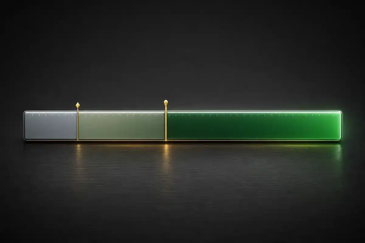
Laboratory reference ranges for ferritin are set primarily to exclude iron deficiency anaemia. Most laboratory lower limits sit around 12–15 µg/L. Above that, a result is classified as within range — which, for the purpose of anaemia screening, it is. But those reference ranges were not derived with hair follicle biology in mind; they reflect the threshold below which the body’s oxygen-carrying capacity begins to be compromised, not the threshold below which a hair follicle may begin to struggle.
A substantial body of trichology literature places a different and higher reference point on the ferritin question. In a comprehensive 2006 review published in the Journal of the American Academy of Dermatology, Trost, Bergfeld, and Calogeras examined the diagnostic and clinical relationship between iron deficiency and hair loss across multiple alopecia types, making the case that serum ferritin provides the most reliable estimate of iron stores in this clinical context.4 Earlier clinical work by Rushton and Ramsay found that maintaining adequate serum ferritin levels was a clinically significant factor in women being treated for diffuse androgen-related hair loss, even during active therapeutic management.2
The precise functional threshold discussed in the trichology literature is not settled — figures ranging from 30 µg/L to 70 µg/L appear across different studies and clinical guidelines, reflecting the absence of a single agreed standard. But even taking the lower end of this range, the gap between the laboratory anaemia cutoff (≥12–15 µg/L) and the trichology reference point is not marginal. A ferritin result of 20 or 25 µg/L passes the anaemia screen comfortably. Whether it is adequate for sustained hair follicle function is a different question — and one the standard panel is not designed to answer.
In a large population study of 5,110 women aged 35–60, Deloche and colleagues found in 2007 that 59% of those reporting excessive hair loss had ferritin below 40 µg/L — a larger proportion than in the moderate or no-hair-loss groups in the same cohort.5 The study relied on self-reported shedding rather than clinical diagnosis, and the association does not establish causation. But at that sample size, it functions as a meaningful epidemiological signal.
The biology of a hungry follicle
To understand why the threshold gap matters, it helps to understand what iron is actually doing inside a growing hair follicle — because the relationship is not simply correlational. There is a clear biological mechanism.
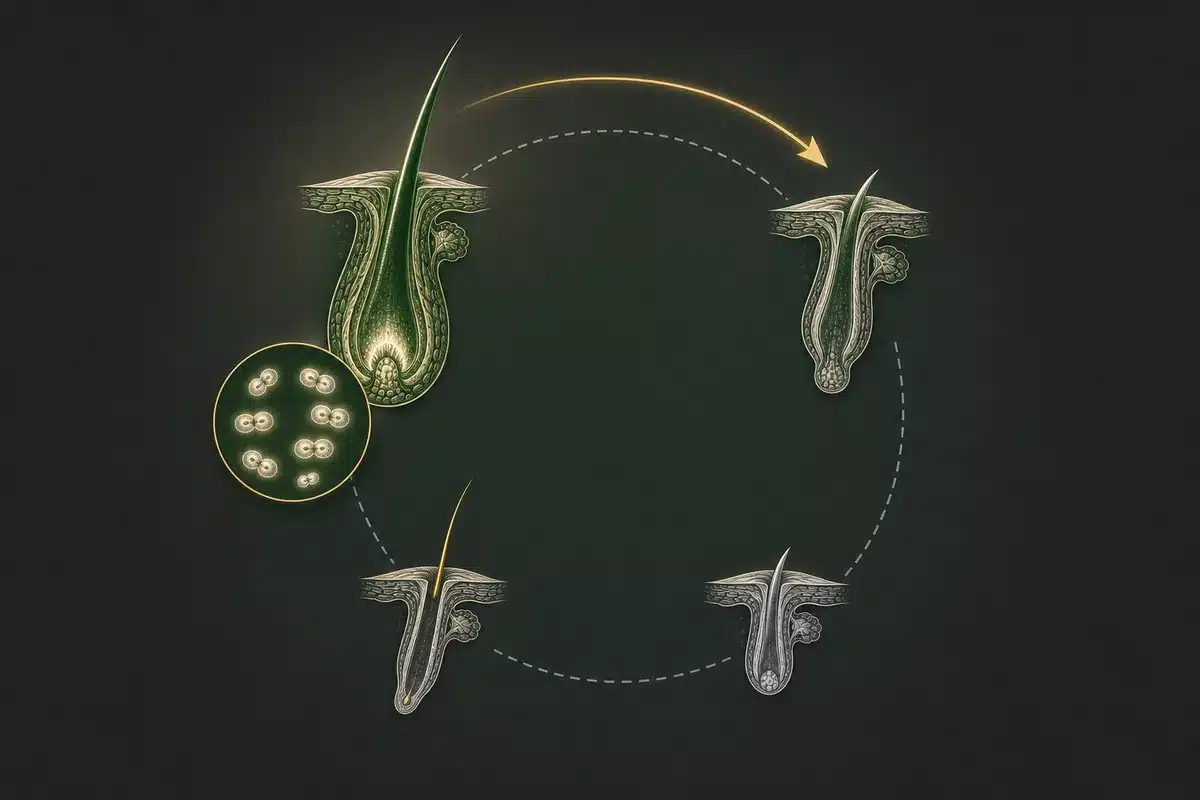
During the anagen phase — the active growth period that lasts two to seven years — the matrix cells at the base of the hair bulb divide continuously. They are among the most rapidly proliferating cells in the adult human body, generating the keratin that builds the hair shaft upward. That rate of division demands a constant and substantial supply of newly synthesised DNA, which requires deoxyribonucleotides, which are in turn produced by an enzyme called ribonucleotide reductase.
Ribonucleotide reductase is iron-dependent: it requires iron as a functional co-factor to catalyse the conversion of ribonucleotides to the deoxyribonucleotides needed for DNA replication. Research by Furukawa and colleagues demonstrated directly that iron deprivation reduces ribonucleotide reductase activity, producing measurable downstream decreases in DNA synthesis and cell proliferation.1 When iron stores fall, this enzymatic pathway operates below capacity.
The follicle does not receive a protected allocation of iron. It competes with other tissues for whatever is available from ferritin reserves. When those reserves are low, iron is directed preferentially toward oxygen-carrying functions — this is biologically rational, but it means the hair follicle, important but not acutely life-sustaining, is among the first structures to experience a shortfall. The consequence, accumulating over weeks to months, may be premature transition out of anagen: follicles entering the resting telogen phase earlier than their biology would otherwise dictate, with the characteristic diffuse shedding following two to three months later.
What the evidence actually shows
The research on ferritin and hair loss is genuinely mixed, and that matters for how the evidence should be read and communicated.
Kantor, Kessler, Brooks, and Cotsarelis found in a 2003 cross-sectional study that women with androgenetic alopecia had significantly lower mean serum ferritin levels compared to controls without hair loss — a statistically meaningful difference at the population level.3 For alopecia areata, a similar pattern was observed. For telogen effluvium, a difference was present in the data but did not reach statistical significance in their sample.
A more complex finding came from a large controlled study by Olsen and colleagues at the Duke University Hair Disorders Clinic in 2010, which examined 381 women with female pattern hair loss or chronic telogen effluvium alongside 76 controls. Using ferritin ≤40 µg/L as the threshold, iron deficiency was found in 58.8% of the female pattern hair loss group and 63.8% of the chronic telogen effluvium group — but also in 72.3% of the control women with no hair loss.6 The difference between the hair loss and control groups was not statistically significant. The study’s conclusion was that iron deficiency at this threshold is extremely common in premenopausal women regardless of hair loss status — meaning low ferritin is not a reliable independent differentiator between those with and without hair shedding.
What this means in practice: low ferritin does not automatically explain every case of diffuse hair loss, and a single ferritin measurement — whatever the result — provides a limited picture. The more clinically meaningful approach is tracking ferritin over time, alongside scalp health observations and other biomarker data, to identify longitudinal patterns that a single snapshot cannot reveal. Individual trajectory matters more than a single reading.
The conversation worth having
If you have been told your blood results are normal after raising concerns about hair shedding, the following are worth putting directly to your GP or dermatologist:
- Was ferritin specifically included in the panel, or only haemoglobin?
- If ferritin was measured, what is the actual numerical result — not just the pass or fail designation?
- Where does that value sit relative to the thresholds discussed in trichology research, and would longitudinal monitoring be appropriate?
None of this is self-diagnosis. Whether addressing iron stores is clinically appropriate — and through what means — is a decision that belongs with your GP, guided by your complete clinical picture. But entering that conversation with the actual ferritin number in hand, and an understanding of why the laboratory normal range may not be the most relevant reference point for a hair loss question, changes the quality of the discussion that is possible.
Ferritin can shift meaningfully over months, influenced by diet, absorption, menstrual blood loss, or underlying health changes. A measurement taken once, filed, and never revisited is a single data point. A value tracked longitudinally alongside scalp health observations — shedding, density changes, photographs taken the same way each time — begins to tell a substantively more useful story, and provides a more productive foundation for any clinical intervention discussion.
If you have a recent ferritin result, you can enter it in Scalp Score, free with no sign-up, and see it beside the ranges in the published literature, with the source for each range. The value itself is never sent for an analysis, and it is stored on your phone unless you turn on the backup. Discuss the implications with your GP or dermatologist — the number is the starting point, not the answer.
References
- Furukawa T, Naitoh Y, Kohno H, Tokunaga R, & Taketani S. (1992). Iron deprivation decreases ribonucleotide reductase activity and DNA synthesis. Life Sciences, 50(26), 2059–2065. Read study
- Rushton DH, & Ramsay ID. (1992). The importance of adequate serum ferritin levels during oral cyproterone acetate and ethinyl oestradiol treatment of diffuse androgen-dependent alopecia in women. Clinical Endocrinology, 36(4), 421–427. Read study
- Kantor J, Kessler LJ, Brooks DG, & Cotsarelis G. (2003). Decreased serum ferritin is associated with alopecia in women. Journal of Investigative Dermatology, 121(5), 985–988. Read study
- Trost LB, Bergfeld WF, & Calogeras E. (2006). The diagnosis and treatment of iron deficiency and its potential relationship to hair loss. Journal of the American Academy of Dermatology, 54(5), 824–844. Read study
- Deloche C, Bastien P, Chadoutaud S, Galan P, Bertrais S, Hercberg S, & de Lacharrière O. (2007). Low iron stores: a risk factor for excessive hair loss in non-menopausal women. European Journal of Dermatology, 17(6), 507–512. Read study
- Olsen EA, Reed KB, Cacchio PB, & Caudill L. (2010). Iron deficiency in female pattern hair loss, chronic telogen effluvium, and control groups. Journal of the American Academy of Dermatology, 63(6), 991–999. Read study